Mohs Micrographic Surgery in Anderson, Seneca, Abbeville, Pendleton and Clemson, SC
Mohs Micrographic Surgery is a very specialized surgical technique for the removal of skin cancers. During the procedure, thin layers of skin are progressively removed until all the skin cancer has been removed. If they detect any cancer cells on the margins of the tissue, the surgeon will know exactly where the cells are located and can remove another layer of tissue from that precise location. The doctor then repeats the process until no cancer cells are detected. Mohs surgery is performed as an outpatient procedure in the physician’s office. The surgeon acts as a pathologist and reconstructive surgeon in order to complete the Mohs micrographic surgery during one patient visit. This unique procedure offers the best cure rate and allows the surgeon to save as much normal, healthy tissue as possible.
As a result, this leads to faster recovery times and less scarring. This can be especially useful for larger skin cancers and on certain parts of the body such as the eyelids, nose, and ears.
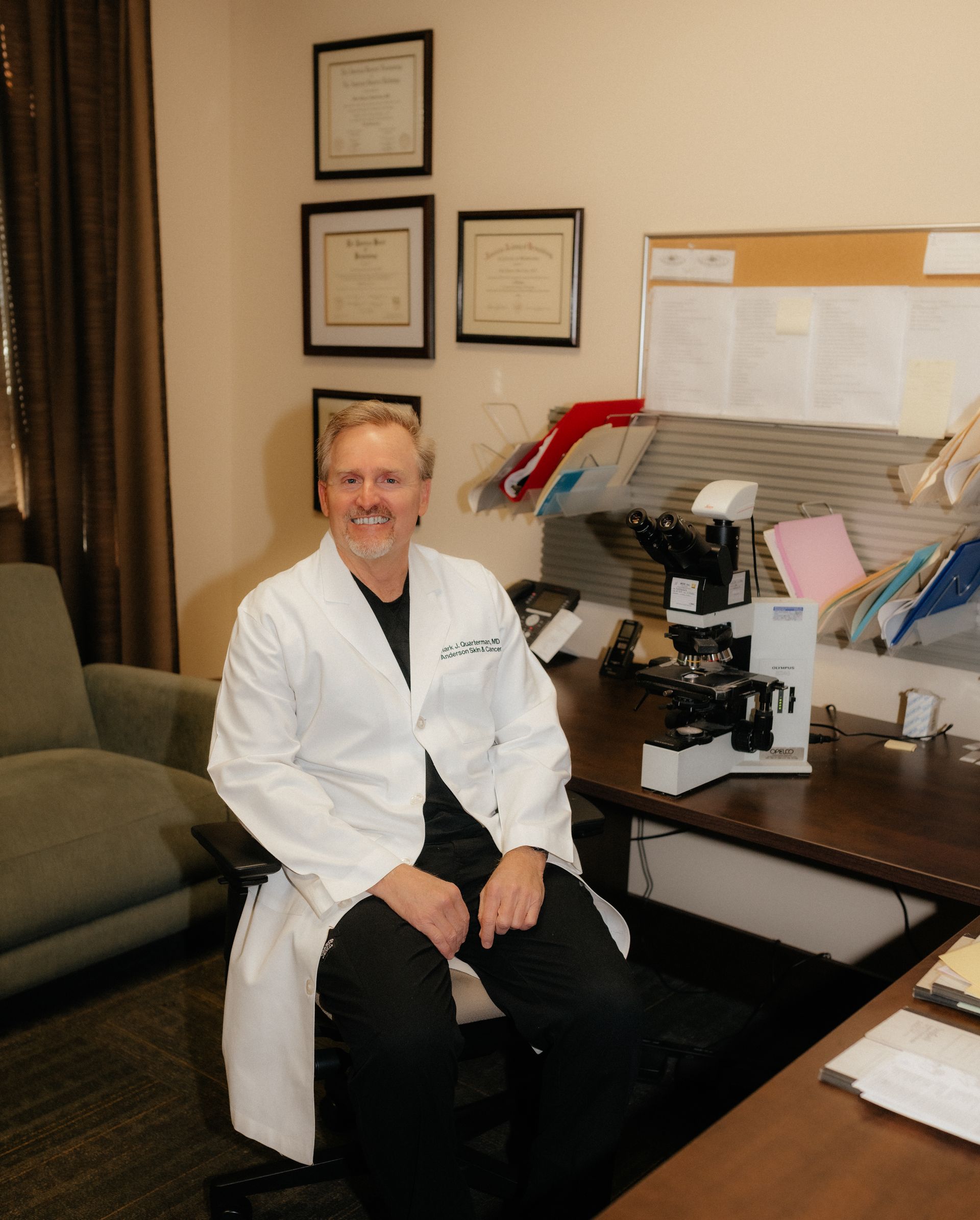

Board Certified in Micrographic Dermatologic Surgery
Dr. Mark Quarterman is board certified in Micrographic Dermatologic Surgery and has performed many MOHS surgeries in the past 25 years.
Understanding the MOHS Procedure
The area to be treated is examined, cleaned, and injected with a local anesthetic to numb the area. The Mohs surgeon removes visible cancer, along with a thin layer of additional normal tissue. The patient waits while the tissue is being processed and examined. The Mohs surgeon carefully examines the entire undersurface and complete edge of the specimen. Upon microscopic examination, if residual cancer is found, the Mohs surgeon removes additional tissue. This process is repeated as many times as necessary to remove any remaining cancerous areas within the tissue specimen. When microscopic examination reveals that there is no remaining cancer, the surgical defect is ready for repair.